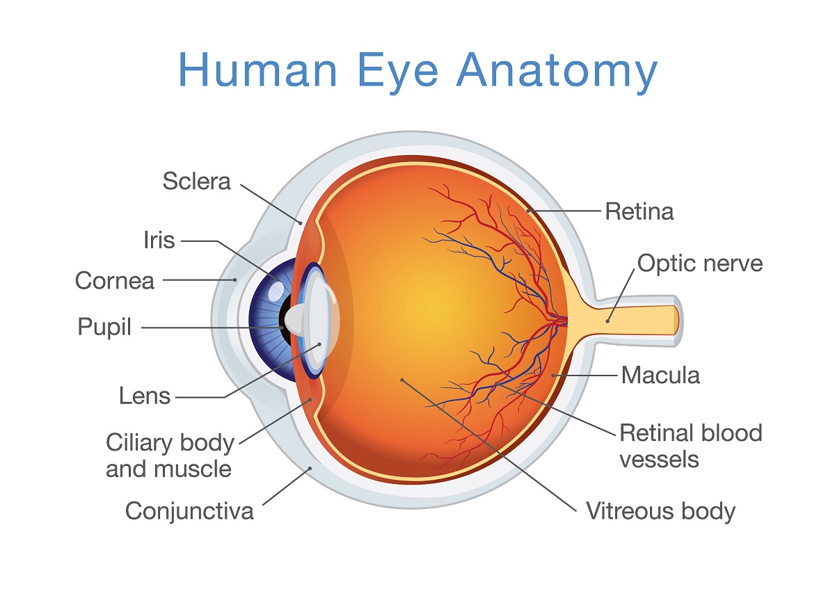
The eye is a fluid-filled sphere enclosed by three layers of tissue Figure 111. These muscles work to rotate the eye and move the.
 |
| Anatomy Of A Normal Human Eye Amdf |
The iris Ciliary body Choroid.

. The course aims through online learning and completion of tests to help you develop and improve your knowledge of the anatomy of the eye. Bony cavity within the skull that houses the eye and its associated structures muscles of the eye eyelid periorbital fat lacrimal apparatus Bones of the orbit. In order for eyes to be able to see the cornea and the lens must refract the light entering the eyes and focus it on the retina which is the layer of light sensitive cells lining. Anatomy of the eye Videos Flashcards High Yield Notes Practice Questions.
The teaching provided by Gloucestershire. Anatomy of the eye. At the front of. WebMDs Eyes Anatomy Pages provide a detailed picture and definition of the human eyes.
Learn and reinforce your understanding of Anatomy of the eye. Most of the outer layer is composed of a tough white fibrous tissue the sclera. - Osmosis is an efficient enjoyable and. It is made of a dense strong fibrous wall consisting of the sclera that is 56 th and the cornea that is anterior 16 th of the eyeball.
Human eyes function when the three parts of the eye work together to let in light and focus the light to the retina which sends electrical signals through the optic nerve to the brain. The retina connects to the optic nerve to send vision. Anatomy of the Eye. From front to back the key anatomy of the eye includes the tear film cornea anterior chamber iris lens vitreous and retina.
There are six extraocular muscles that attach to the outside of the eye from the bone in the eyes socket. A closer look at the parts of the eye. When surveyed about the five senses sight hearing taste smell and touch people consistently report that their eyesight. It makes up the outermost part of eye anatomy.
Learn about their function and problems that can affect the eyes. It is located underneath the white part of the eye the sclera and is composed of three parts.
 |
| 0514 Human Eye Anatomy Medical Images For Powerpoint Powerpoint Presentation Images Templates Ppt Slide Templates For Presentation |
 |
| How Your Eye Works Parts Of The Eye Look After Your Eyes |
 |
| 20 424 Eye Anatomy Stock Photos Pictures Royalty Free Images Istock |
 |
| Human Eye Anatomy Infographic Lifemap Discovery |
 |
| Laminated Eye Anatomical Poster Human Eye Anatomy Chart 18 X 27 Amazon In Industrial Scientific |